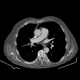
Aufnahme 1
Lungenarterienembolie
Klinik:
79-jähriger Patient mit Z.n. Zystektomie bei Blasen-Ca. Aktuell keine Beschwerden.
Untersuchung/Befund:
Als Zufallsbefund zeigte sich in der Kontrastmittel-gestützten CT- Untersuchung des Thorax eine bilaterale Lungenarterienembolie mit Kontrastmittelaussparungen in der proximalen Arteria pulmonalis beidseits sowie auf Segment- und Subsegmentniveau (Bild 7-1 und 7-2, Pfeile). Nebenbefundlich zeigt sich eine kalzifizierende Arteriosklerose.
Beurteilung:
Bild einer bilateralen subakuten Lungenarterien-Embolie (LAE).
Epidemiologie
Allgemein gilt die Virchow’sche Trias als Risikofaktoren (Veränderung der Blutzusammensetzung durch Medikamente oder erbliche Faktoren, Veränderung der Gefäße durch Verletzungen oder Entzündungen, verminderte Strömungsgeschwindigkeit durch Krampfadern oder Bettlägerigkeit).
Daraus ergeben sich folgende Risikofaktoren für die Begünstigung der Bildung einer Lungenarterienembolie:
- Primäre Hyperkoagulablitäts-Syndrome wie z.B. Protein-C-Mangel, Protein-S-Mangel, Antithrombin III-Mangel, Lupus-Antikoagulans, Faktor-V-Leiden
- Postoperativ (nach einer stattgehabten Operation) aufgrund von längerer Bettruhe und /oder Immobilität
- Malignität (Krebserkrankungen) als paraneoplastische Veränderung
- HIV/AIDS: 2-10-fach erhöhtes Risiko
- COVID-19
- Medikamente wie z.B. orale Kontrazeptiva (Verhütungsmittel)
- Schwangerschaft
- Bekannte oder frühere Tiefe-Beinvenen-Thrombose (TVT)
Pathologie/Labor:
D-Dimere wird bei Patient:innen bei V. a. Lungenarterienembolie bestimmt. Ein normales D-Dimer hat einen negativen Vorhersagewert von fast 100 % (schließt eine PE praktisch aus), sodass weitere Tests nicht erforderlich sind. Erhöhte D-Dimere wiederum treten bei einer Lungenembolie auf, können aber auch viele andere Ursachen haben und sind daher unspezifisch. So sollten hier weitere Abklärungen erfolgen.
Im Gegensatz zur akuten Lungenembolie handelt es sich bei chronischen Thrombembolien häufig um vollständige Verschlüsse oder nicht okklusive Füllungsdefekte in der Peripherie des betroffenen Gefäßes, die stumpfe Winkel mit der Gefäßwand bilden. Der Thrombus kann verkalkt sein.
Therapie/Prognose:
Die erste Behandlung ist die kardiopulmonale Unterstützung. Bei Patienten, bei denen keine aktive Blutung droht, wird eine Antikoagulation durchgeführt. Wenn die Embolien groß sind oder eine hohe Thrombuslast vorliegt, ist eine Thrombolyse eine Option. In einigen Fällen ist eine Embolektomie oder das Einsetzen eines Vena-Cava-Filters erforderlich. Das rechtsventrikuläre Versagen aufgrund von Drucküberlastung gilt als die Haupttodesursache bei schwerer Lungenembolie.
Differentialdiagnose:
- Artefakte: Artefakt der Blutströmung, Kontrast-Blutspiegel (durch langsamen Fluss), Atembewegung, Strahlenverhärtung, medizinische Geräte, z. B. Katheter, orthopädische Prothesen, Arme des Patienten / der Patientin in gesenkter Position, Bewegung der Patient:innen usw.
- Iatrogen: kavopulmonale Anastomose
- Neoplastisch: Sarkom der Pulmonalarterie
- Entzündlich: Vaskulitis der Lungenarterie, z. B. Takayasu-Arteriitis
- Verwechslung von chronischen und akuten Embolien
Literatur
- Worsley D, Alavi A, Aronchick J, Chen J, Greenspan R, Ravin C. Chest Radiographic Findings in Patients with Acute Pulmonary Embolism: Observations from the PIOPED Study. Radiology. 1993;189(1):133-6.
- Corwin M, Donohoo J, Partridge R, Egglin T, Mayo-Smith W. Do Emergency Physicians Use Serum D-Dimer Effectively to Determine the Need for CT when Evaluating Patients for Pulmonary Embolism? Review of 5,344 Consecutive Patients. AJR Am J Roentgenol. 2009;192(5):1319-23.
- Han D, Lee K, Franquet T et al. Thrombotic and Nonthrombotic Pulmonary Arterial Embolism: Spectrum of Imaging Findings. Radiographics. 2003;23(6):1521-39.
- Stein P, Woodard P, Weg J et al. Diagnostic Pathways in Acute Pulmonary Embolism: Recommendations of the PIOPED II Investigators. Radiology. 2007;242(1):15-21.
- Ghaye B, Ghuysen A, Bruyere P, D'Orio V, Dondelinger R. Can CT Pulmonary Angiography Allow Assessment of Severity and Prognosis in Patients Presenting with Pulmonary Embolism? What the Radiologist Needs to Know. Radiographics. 2006;26(1):23-39; discussion 39.
- Ocak I & Fuhrman C. CT Angiography Findings of the Left Atrium and Right Ventricle in Patients with Massive Pulmonary Embolism. AJR Am J Roentgenol. 2008;191(4):1072-6.
- Castañer E, Gallardo X, Ballesteros E et al. CT Diagnosis of Chronic Pulmonary Thromboembolism. Radiographics. 2009;29(1):31-50; discussion 50.
- Kang D, Thilo C, Schoepf U et al. CT Signs of Right Ventricular Dysfunction: Prognostic Role in Acute Pulmonary Embolism. JACC Cardiovasc Imaging. 2011;4(8):841-9.
- Wittram C, Maher M, Yoo A, Kalra M, Shepard J, McLoud T. CT Angiography of Pulmonary Embolism: Diagnostic Criteria and Causes of Misdiagnosis. Radiographics. 2004;24(5):1219-38.
- Rossi S, Goodman P, Franquet T. Nonthrombotic Pulmonary Emboli. AJR Am J Roentgenol. 2000;174(6):1499-508.
- Williams J & Wilcox W. Pulmonary Embolism. Roentgenographic and Angiographic Considerations. Am J Roentgenol Radium Ther Nucl Med. 1963;89:333-42.
- Tatco V & Piedad H. The Validity of Hyperdense Lumen Sign in Non-Contrast Chest CT Scans in the Detection of Pulmonary Thromboembolism. Int J Cardiovasc Imaging. 2011;27(3):433-40.
- Stein P, Chenevert T, Fowler S et al. Gadolinium-Enhanced Magnetic Resonance Angiography for Pulmonary Embolism: A Multicenter Prospective Study (PIOPED III). Ann Intern Med. 2010;152(7):434-43, W142-3.
- Konstantinides S, Torbicki A, Agnelli G et al. 2014 ESC Guidelines on the Diagnosis and Management of Acute Pulmonary Embolism. Eur Heart J. 2014;35(43):3033-69, 3069a.
- Gabrielli R, Vitale S, Costanzo A, Carra A. Our Experience of Popliteal Vein Aneurysm. Interact Cardiovasc Thorac Surg. 2010;11(6):835-7.
- Martin L. Gunn. Pearls and Pitfalls in Emergency Radiology. (2013) ISBN: 9781139619899 -
- Palla A, Donnamaria V, Petruzzelli S, Rossi G, Riccetti G, Giuntini C. Enlargement of the Right Descending Pulmonary Artery in Pulmonary Embolism. AJR Am J Roentgenol. 1983;141(3):513-7.
- Chang C & Davis W. A Roentgen Sign of Pulmonary Infarction. Clin Radiol. 1965;16(2):141-7.
- Jaff M, McMurtry M, Archer S et al. Management of Massive and Submassive Pulmonary Embolism, Iliofemoral Deep Vein Thrombosis, and Chronic Thromboembolic Pulmonary Hypertension: A Scientific Statement from the American Heart Association. Circulation. 2011;123(16):1788-830.
- Aghayev A, Furlan A, Patil A et al. The Rate of Resolution of Clot Burden Measured by Pulmonary CT Angiography in Patients with Acute Pulmonary Embolism. AJR Am J Roentgenol. 2013;200(4):791-7.
- Stein P, Yaekoub A, Matta F et al. Resolution of Pulmonary Embolism on CT Pulmonary Angiography. AJR Am J Roentgenol. 2010;194(5):1263-8.
- Bibas M, Biava G, Antinori A. HIV-Associated Venous Thromboembolism. Mediterr J Hematol Infect Dis. 2011;3(1):e2011030.
- Vallianou N, Lazarou V, Tzangarakis J, Barounis R, Sioula E. Pulmonary Embolism as the First Manifestation of Multiple Myeloma. Case Rep Med. 2013;2013:236913.
- Fields J, Davis J, Girson L et al. Transthoracic Echocardiography for Diagnosing Pulmonary Embolism: A Systematic Review and Meta-Analysis. J Am Soc Echocardiogr. 2017;30(7):714-723.e4.
- Kosuge M, Kimura K, Ishikawa T et al. Electrocardiographic Differentiation Between Acute Pulmonary Embolism and Acute Coronary Syndromes on the Basis of Negative T Waves. Am J Cardiol. 2007;99(6):817-21.
- Kosuge M, Ebina T, Hibi K et al. Differences in Negative T Waves Among Acute Coronary Syndrome, Acute Pulmonary Embolism, and Takotsubo Cardiomyopathy. Eur Heart J Acute Cardiovasc Care. 2012;1(4):349-57.
- Danzi G, Loffi M, Galeazzi G, Gherbesi E. Acute Pulmonary Embolism and COVID-19 Pneumonia: A Random Association? Eur Heart J. 2020;41(19):1858.
- Pesavento R, Filippi L, Palla A et al. Impact of Residual Pulmonary Obstruction on the Long-Term Outcome of Patients with Pulmonary Embolism. Eur Respir J. 2017;49(5):1601980.
- Moore A, Wachsmann J, Chamarthy M, Panjikaran L, Tanabe Y, Rajiah P. Imaging of Acute Pulmonary Embolism: An Update. Cardiovasc Diagn Ther. 2018;8(3):225-43.
- Asah D, Raju S, Ghosh S, Mukhopadhyay S, Mehta A. Nonthrombotic Pulmonary Embolism From Inorganic Particulate Matter and Foreign Bodies. Chest. 2018;153(5):1249-65.
- Hirsh J. Risk of Thrombosis with Lenalidomide and Its Prevention with Aspirin. Chest. 2007;131(1):275-7.
- Palm V, Rengier F, Rajiah P, Heussel C, Partovi S. Acute Pulmonary Embolism: Imaging Techniques, Findings, Endovascular Treatment and Differential Diagnoses. Rofo. 2020;192(1):38-49.
- Yazdani M, Lau C, Lempel J et al. Historical Evolution of Imaging Techniques for the Evaluation of Pulmonary Embolism:RSNA Centennial Article. Radiographics. 2015;35(4):1245-62.
- Tseng E. Subsegmental Pulmonary Embolism: To Treat or Not to Treat? The Hematologist. 2022;19(2).

Aufnahme 2