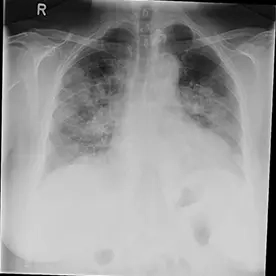
Aufnahme 1
COVID-19 Pneumonie
Klinik:
61-jährige Patientin mit Z. n. Apoplex, fieberhafter bronchopulmonaler Infekt
Untersuchung/Befund:
Es zeigen sich bipulmonale konsolidierende Infiltrate ohne begleitende Pleuraergüsse. Bei positivem Test der Patientin auf COVID-19 ist hier von einer COVID-19-Pneumonie auszugehen.
Beurteilung:
Covid-19 Pneumonie (SARS-CoV-2)
Epidemiologie
Im Dezember 2022 beläuft sich die Zahl der bestätigten COVID-19-Fälle weltweit auf über 600 Millionen. Die R0 (Basisreproduktionszahl) des ursprünglichen Wildtyps von SARS-CoV-2 wurde auf 2,2 bis 3,3 in einer Population geschätzt, d. h. jedes infizierte Individuum verursacht im Durchschnitt 2-3 neue Infektionen. Es sind mehrere Varianten aufgetaucht, und einige Varianten sind stärker übertragbar. Die Inkubationszeit für COVID-19 wurde ursprünglich auf etwa 5 Tage berechnet. Eine große Meta-Analyse von 53 Studien weltweit ergab eine mittlere Inkubationszeit von 6 Tagen. Im Dezember 2022 übersteigt die offizielle Zahl der Todesfälle durch COVID-19 weltweit sechs Millionen. Die Sterblichkeitsrate liegt bei ~2-3%. Es wird vermutet, dass die tatsächliche Sterblichkeitsrate niedriger ist, weil viele milde/asymptomatische Fälle nicht getestet werden, wodurch die scheinbare Sterblichkeitsrate nach oben verzerrt wird. Bis Dezember 2022 waren weltweit mehr als 13 Milliarden Impfstoffdosen verabreicht worden.
Pathologie/Labor:
Ätiologie
Am 9. Januar 2020 bestätigte die Weltgesundheitsorganisation (WHO), dass SARS-CoV-2 die Ursache von COVID-19 war. Es ist einer der beiden Stämme der SARS-CoV-Spezies, von denen bekannt ist, dass sie beim Menschen Krankheiten verursachen, der andere ist das ursprüngliche schwere akute respiratorische Syndrom-Coronavirus (SARS-CoV-1), die Ursache von SARS. Es gehört zur Gattung der ß-Coronaviren, einer der Gattungen der Virusfamilie der Coronaviridae. Coronaviren sind umhüllte einzelsträngige RNA-Viren, die bei Menschen, Säugetieren und Vögeln vorkommen. Diese Viren sind für Lungen-, Leber-, ZNS- und Darmerkrankungen verantwortlich. Wie viele menschliche Infektionen ist auch SARS-CoV-2 zoonotisch. Das der genetischen Sequenz nach nächstgelegene tierische Coronavirus ist ein Fledermaus-Coronavirus, und dieses ist wahrscheinlich der eigentliche Ursprung des Virus. Die Krankheit kann auch durch Schlangen übertragen werden. Es sind sechs weitere Coronaviren bekannt, die beim Menschen Krankheiten verursachen. Zwei davon sind Zoonosen: das Coronavirus des schweren akuten respiratorischen Syndroms (SARS-CoV-1) und das Coronavirus des Mittleren Ostens (MERS-CoV), die tödlich verlaufen können. Die übrigen vier Viren verursachen alle eine simple Erkältungssymptome.
Pathophysiologie
Das SARS-CoV-2-Virus, wie auch die eng verwandten MERS- und SARS-Coronaviren, dringt in die Zellen ein, indem es sein Virion-Spike-Protein (auch S-Protein genannt) an den Angiotensin-Converting-Enzyme-2-Rezeptor (ACE2-Rezeptor) bindet. Dieser Rezeptor findet sich häufig auf den Alveolarzellen des Lungenepithels und ist die Ursache für die Entwicklung von Atemwegssymptomen, die die häufigste Erscheinungsform von COVID-19 50 sind. Es wird vermutet, dass die weniger häufigen kardiovaskulären Wirkungen ebenfalls über denselben ACE2-Rezeptor vermittelt werden, der ebenfalls häufig auf den Zellen des kardiovaskulären Systems exprimiert wird.
Varianten
Das SARS-CoV-2-Virus mutiert wie alle anderen Viren ständig und bringt dabei immer wieder neue Varianten hervor, wobei bestimmte Varianten den Wissenschaftler:innen besondere Sorgen bereiten. Diese könnten besonders übertragbar oder krankmachend sein. Diese wurden von der Weltgesundheitsorganisation (WHO) als "besorgniserregende Varianten" (VOC) bezeichnet. Das "ursprüngliche" Virus, d. h. wie es vor dem Auftreten der Alpha-Variante aussah, wird heute als "Wildtyp"-Virus bezeichnet. Früher gab es mehrere "Varianten von Interesse" (VOI), die genetische Sequenzen aufwiesen, die wichtige virale Eigenschaften beeinflussen konnten, z. B. Übertragbarkeit, Pathogenität, Fähigkeit zur Umgehung von Impfstoffen usw., und bei denen festgestellt wurde, dass sie mehrere Krankheitscluster bilden oder eine erhebliche Übertragung in der Gemeinschaft bewirken, aber keine davon wird derzeit als solche bezeichnet.
RT-PCR-Test
Der diagnostische Standardtest für SARS-CoV-2 ist der Reverse-Transkriptase-Polymerase-Kettenreaktion (RT-PCR)-Test in Echtzeit. Dieser ermöglicht auch die Suche nach spezifischen Sequenzen aus dem viralen Genom: E (Hüllprotein-Gen), N (Nukleokapsid-Protein-Gen) und RdRP (RNA-abhängiges RNA-Polymerase-Gen) 207. Man geht davon aus, dass er hochspezifisch ist, wobei die Sensitivität zwischen 60-70 % 32 und 95-97 % liegt. In einer Meta-Analyse wurde die gepoolte Sensitivität der RT-PCR mit 89 % beziffert. Falsch-negative Ergebnisse sind also ein echtes klinisches Problem, und in einem einzigen Fall können mehrere negative Tests erforderlich sein, um die Krankheit sicher ausschließen zu können.
Laboruntersuchungen
Die häufigsten ergänzenden auffälligen Laborbefunde bei Patient:innen waren wie gefolgt:
- Lymphopenie
- Thrombozytose
- erhöhte Prothrombinzeit (PT)
- erhöhte Laktatdehydrogenase
- leicht erhöhte Entzündungsmarker
- Erhöhte D-Dimere
- leicht erhöhte Serum-Amylase: 17 % der Patienten
(Es wurde über akute Pankreatitis berichtet, wobei unklar ist, ob ein kausaler Zusammenhang mit SARS-CoV-2 oder ein Epiphänomen besteht)
- Leicht erhöhte Alanin-Aminotransferase (ALT) und Aspartat-Aminotransferase (AST) ; der Bilirubinanstieg ist im Allgemeinen leicht erhöht, die Werte alkalische Phosphatase (ALP) und Gamma Glutamyltransferase (GGT) bleiben normalerweise normal.
Therapie/Prognose:
Die meisten Ressourcen wurden auf Maßnahmen des öffentlichen Gesundheitswesens konzentriert, um eine weitere Übertragung des Virus auf andere Menschen zu verhindern; dazu gehören:
- gründliches Händewaschen
- Tragen von Gesichtsmasken
- Distanzierung (Social distancing)
- Meiden großer Menschenmengen/überfüllter Umgebungen
- Selbstisolierung
- Bessere Belüftung, insbesondere in öffentlichen Innenräumen, an Arbeitsplätzen, in Bildungseinrichtungen, in Gesundheitseinrichtungen und in Gemeinschaftswohnheimen für ältere Menschen
In Gesundheitseinrichtungen sind eine schnelle Diagnose notwendig, um die betroffenen in Quarantäne zu stellen und wirksame unterstützende Therapien anzubieten. Dies umfasst empirische Behandlungen mit Antibiotika, Virostatika und unterstützenden Maßnahmen. Mechanische Beatmung, sowohl invasiv als auch nicht-invasiv, und extrakorporale Membranoxygenierung (ECMO) wurden ebenfalls eingesetzt, wenn dies klinisch notwendig war.
Impfstoffe
Das primäre Ziel bei der Entwicklung von Impfstoffen gegen Coronaviren war das Spike-Protein (S-Protein), das sich auf der Oberfläche des Virionpartikels befindet und in vivo das wichtigste Antigen für die Auslösung einer Immunreaktion ist. Humanimpfstoffe gegen Coronaviren werden seit dem SARS-Ausbruch 2002-2004 entwickelt, aber keiner wurde bisher für die Immunisierung gegen SARS oder MERS zugelassen.
Impfstoffe gegen SARS-CoV-2 können nach ihren unterschiedlichen Wirkmechanismen klassifiziert werden:
- genetische Impfstoffe z. B. Pfizer
- adenovirale Vektoren, z. B. AstraZeneca
- inaktiviertes Virion, z. B. Sinopharm
- Untereinheit z.B. Novavax
Long Covid
Der Begriff "Long Covid" wurde verwendet, um den Zustand zu bezeichnen, bei dem Personen, die sich von COVID-19 erholt haben, immer noch an anhaltenden Symptomen leiden, oder für Personen mit COVID-19 über einen längeren Zeitraum an Symptomen leiden als normal.
Ab September 2021 gibt es immer mehr veröffentlichte Belege für die Langzeitfolgen einer COVID-19-Infektion. Zu den häufigsten Symptomen gehören chronische Müdigkeit, Atemnot, Gelenkschmerzen und Brustbeschwerden.
Differentialdiagnose:
- Andere virale Lungenentzündung einschließlich:
Influenzapneumonie A und B
Verteilung mehr entlang der bronchovaskulären Bündel
Verdickung der Bronchialwände
- Paramyxovirus-Pneumonie
- Zytomegalievirus (CMV)-Pneumonie
- Adenovirus-Pneumonie 71,72
- SARS-CoV-Pneumonie
- MERS-Coronavirus
- HSV-Pneumonie
oft Pleuraergüsse als Mittreaktion
Vor allem bei Kinder und Heranwachsenden Lungenentzündung durch das Respiratorische Synzytialvirus (RSV), sowie bei Immungeschwächten atypische bakterielle Lungenentzündung wie z.B. Mykoplasmen-Pneumonie
- Aspirationspneumonitis
Literatur
- Zhu N, Zhang D, Wang W et al. A Novel Coronavirus from Patients with Pneumonia in China, 2019. N Engl J Med. 2020;382(8):727-33.
- Perlman S. Another Decade, Another Coronavirus. N Engl J Med. 2020;382(8):760-2.
- Huang C, Wang Y, Li X et al. Clinical Features of Patients Infected with 2019 Novel Coronavirus in Wuhan, China. Lancet. 2020;395(10223):497-506
- Douglas D. Richman, Richard J. Whitley, Frederick G. Hayden. Clinical Virology. (2016) ISBN: 9781555819422
- COVID-19 Dashboard by the Center for Systems Science and Engineering (CSSE) at Johns Hopkins University (JHU)
- Chen N, Zhou M, Dong X et al. Epidemiological and Clinical Characteristics of 99 Cases of 2019 Novel Coronavirus Pneumonia in Wuhan, China: A Descriptive Study. Lancet. 2020;395(10223):507-13.
- "WHO Director-General's opening remarks at the media briefing on COVID-19 - 11 March 2020". Who.int, 2020.
- Wu J, Leung K, Leung G. Nowcasting and Forecasting the Potential Domestic and International Spread of the 2019-NCoV Outbreak Originating in Wuhan, China: A Modelling Study. The Lancet. 2020;395(10225):689-97.
- Holshue M, DeBolt C, Lindquist S et al. First Case of 2019 Novel Coronavirus in the United States. N Engl J Med. 2020;382(10):929-36.
- Hui D, I Azhar E, Madani T et al. The Continuing 2019-NCoV Epidemic Threat of Novel Coronaviruses to Global Health - The Latest 2019 Novel Coronavirus Outbreak in Wuhan, China. Int J Infect Dis. 2020;91:264-6.
- Chen Y, Liu Q, Guo D. Emerging Coronaviruses: Genome Structure, Replication, and Pathogenesis. J Med Virol. 2020;92(4):418-23.
- Li Q, Guan X, Wu P et al. Early Transmission Dynamics in Wuhan, China, of Novel Coronavirus-Infected Pneumonia. N Engl J Med. 2020;382(13):1199-207.
- Wang D, Hu B, Hu C et al. Clinical Characteristics of 138 Hospitalized Patients With 2019 Novel Coronavirus-Infected Pneumonia in Wuhan, China. JAMA. 2020;323(11):1061-9.
- Wu P, Hao X, Lau E et al. Real-Time Tentative Assessment of the Epidemiological Characteristics of Novel Coronavirus Infections in Wuhan, China, as at 22 January 2020. Euro Surveill. 2020;25(3).
- WHO 2020, "We now have a name for the #2019nCoV disease: COVID-19. I’ll spell it: C-O-V-I-D hyphen one nine – COVID-19", Tweet, 11 February, viewed 11 February 2020, https://twitter.com/WHO/status/1227248333871173632
- Gorbalenya A, Baker S, Baric R et al. Severe Acute Respiratory Syndrome-Related Coronavirus: The Species and Its Viruses – a Statement of the Coronavirus Study Group. BioRxiv. 2020;:2020.02.07.937862.
- Pan F, Ye T, Sun P et al. Time Course of Lung Changes at Chest CT During Recovery from Coronavirus Disease 2019 (COVID-19). Radiology. 2020;295(3):715-21.
- Velavan T & Meyer C. The COVID-19 Epidemic. Trop Med Int Health. 2020;25(3):278-80.
- Heymann D, Shindo N, Shindo N. COVID-19: What is Next for Public Health? Lancet. 2020;395(10224):542-5.
- Zhang L & Liu Y. Potential Interventions for Novel Coronavirus in China: A Systematic Review. J Med Virol. 2020;92(5):479-90.
- Chen H, Guo J, Wang C et al. Clinical Characteristics and Intrauterine Vertical Transmission Potential of COVID-19 Infection in Nine Pregnant Women: A Retrospective Review of Medical Records. The Lancet. 2020;395(10226):809-15.
- Lim J, Jeon S, Shin H et al. Case of the Index Patient Who Caused Tertiary Transmission of COVID-19 Infection in Korea: The Application of Lopinavir/Ritonavir for the Treatment of COVID-19 Infected Pneumonia Monitored by Quantitative RT-PCR. J Korean Med Sci. 2020;35(6):e79.
- Pan Y, Guan H, Zhou S et al. Initial CT Findings and Temporal Changes in Patients with the Novel Coronavirus Pneumonia (2019-NCoV): A Study of 63 Patients in Wuhan, China. Eur Radiol. 2020;30(6):3306-9.
- [The Epidemiological Characteristics of an Outbreak of 2019 Novel Coronavirus Diseases (COVID-19) in China]. Zhonghua Liu Xing Bing Xue Za Zhi. 2020;41(2):145-51.
- Ng LFP, Hiscox JA. Coronaviruses in animals and humans. (2020) BMJ (Clinical research ed.). 368: m634.
- Shi H, Han X, Jiang N et-al. (2020) Radiological findings from 81 patients with COVID-19 pneumonia in Wuhan, China: a descriptive study. [online] thelancet.com 24 February 2020.
- Lee EYP, Ng M-Y, Khong P-L. (2020) COVID-19 pneumonia: what has CT taught us? [online] thelancet.com. Published: February 24, 2020.
- Wang M, Cao R, Zhang L et-al. Remdesivir and chloroquine effectively inhibit the recently emerged novel coronavirus (2019-nCoV) in vitro. (2020) Cell research
- ClinicalTrials.gov [Internet]. Kalil A: National Institute of Allergy and Infectious Diseases (NIAID) (US). 2020 Feb 21 - . Identifier NCT04280705, A Multicenter, Adaptive, Randomized Blinded Controlled Trial of the Safety and Efficacy of Investigational Therapeutics for the Treatment of COVID-19 in Hospitalized Adults.
- Min Wei, Jingping Yuan, Yu Liu et-al. Novel Coronavirus Infection in Hospitalized Infants Under 1 Year of Age in China. (2020) JAMA.
- Jeffrey P Kanne, Brent P Little, Jonathan H Chung et-al. Essentials for Radiologists on COVID-19: An Update—Radiology Scientific Expert Panel. (2020) Radiology.
- Liu Y, Gayle AA, Wilder-Smith A et-al. The reproductive number of COVID-19 is higher compared to SARS coronavirus. (2020) Journal of travel medicine.
- Tao Ai, Zhenlu Yang, Hongyan Hou et-al. Correlation of Chest CT and RT-PCR Testing in Coronavirus Disease 2019 (COVID-19) in China: A Report of 1014 Cases. (2020) Radiology.
- Lan Lan, Dan Xu, Guangming Ye et-al. Positive RT-PCR Test Results in Patients Recovered From COVID-19. (2020) JAMA.
- Wei Zhao, Zheng Zhong, Xingzhi Xie et-al. Relation Between Chest CT Findings and Clinical Conditions of Coronavirus Disease (COVID-19) Pneumonia: A Multicenter Study. (2020) American Journal of Roentgenology.
- World Health Organization (WHO). WHO Statement Regarding Cluster of Pneumonia Cases in Wuhan, China. Beijing: WHO; 9 January 2020. [Accessed 5 March 2020]. https://www.who.int/china/news/detail/09-01-2020-who-statement-regarding-cluster-of-pneumonia-cases-in-wuhan-china
- Jiang S, Shi Z, Shu Y et-al. A distinct name is needed for the new coronavirus. (2020) Lancet (London, England).
- "World Health Organization: Rational use of personal protective equipment for coronavirus disease 2019 (COVID-19)", 2020.
- Kooraki S, Hosseiny M, Myers L, Gholamrezanezhad A. Coronavirus (COVID-19) Outbreak: What the Department of Radiology Should Know. (2020) Journal of the American College of Radiology : JACR.
- Goh S. Rapid response to "Covid-19: a puzzle with many missing pieces". 2020. BMJ 2020;368:m627
- Qin C, Liu F, Yen TC et-al. F-FDG PET/CT findings of COVID-19: a series of four highly suspected cases. (2020) European journal of nuclear medicine and molecular imaging.
- Xiao F, Tang M, Zheng X et-al. Evidence for gastrointestinal infection of SARS-CoV-2. (2020) Gastroenterology.
- "WHO Director-General's opening remarks at the media briefing on COVID-19 - 11 March 2020". Who.int. 2020.
- "Naming the coronavirus disease (COVID-19) and the virus that causes it". Who.int. 2020.
- The species Severe acute respiratory syndrome-related coronavirus : classifying 2019-nCoV and naming it SARS-CoV-2. (2020) Nature Microbiology.
- Caselli D, Aricò M. 2019-nCoV: Polite with children!. (2020) Pediatric reports. 12 (1): 8495.
- Wei Li, Huaqian Cui, Kunwei Li et-al. Chest computed tomography in children with COVID-19 respiratory infection. Pediatric Radiology.
- Puja Mehta, Daniel F McAuley, Michael Brown et-al. COVID-19: consider cytokine storm syndromes and immunosuppression. Published 13 March 2020 link(20)30628-0. Lancet.com.
- Zheng YY, Ma YT, Zhang JY et-al. COVID-19 and the cardiovascular system. (2020) Nature reviews. Cardiology.
- Bai HX, Hsieh B, Xiong Z et-al. Performance of radiologists in differentiating COVID-19 from viral pneumonia on chest CT. (2020) Radiology.
- “ACR Recommendations for the Use of Chest Radiography and Computed Tomography (CT) for Suspected COVID-19 Infection.” American College of Radiology, 11 Mar. 2020,
- Lu H, Stratton CW, Tang YW. Outbreak of pneumonia of unknown etiology in Wuhan, China: The mystery and the miracle. (2020) Journal of medical virology. 92 (4): 401-402.
- Parr J. Pneumonia in China: lack of information raises concerns among Hong Kong health workers. (2020) BMJ (Clinical research ed.). 368: m56.
- Qian-Yi Peng, Xiao-Ting Wang, Li-Na Zhang. Findings of lung ultrasonography of novel corona virus pneumonia during the 2019–2020 epidemic. (2020) Intensive Care Medicine.
- Mahmud Mossa-Basha, Carolyn C Meltzer, Danny C Kim et-al. Radiology Department Preparedness for COVID-19: Radiology Scientific Expert Panel. (2020) Radiology.
- Neeltje van Doremalen, Trenton Bushmaker, Dylan H. Morris et-al. Aerosol and Surface Stability of SARS-CoV-2 as Compared with SARS-CoV-1. (2020) New England Journal of Medicine.
- Dong Y, Mo X, Hu Y et-al. Epidemiological Characteristics of 2143 Pediatric Patients With 2019 Coronavirus Disease in China. (2020) Pediatrics.
- Amici C, Di Caro A, Ciucci A et-al. Indomethacin has a potent antiviral activity against SARS coronavirus. (2006) Antiviral therapy. 11 (8): 1021-30.
- Wu C, Chen X, Cai Y et-al. Risk Factors Associated With Acute Respiratory Distress Syndrome and Death in Patients With Coronavirus Disease 2019 Pneumonia in Wuhan, China. (2020) JAMA internal medicine.
- Erika Poggiali, Alessandro Dacrema, Davide Bastoni et-al. Can Lung US Help Critical Care Clinicians in the Early Diagnosis of Novel Coronavirus (COVID-19) Pneumonia?. (2020) Radiology.
- Stefania Ianniello, Claudia Lucia Piccolo, Grazia L Buquicchio et-al. First-line diagnosis of paediatric pneumonia in emergency: lung ultrasound (LUS) in addition to chest-X-ray (CXR) and its role in follow-up. (2016) The British Journal of Radiology. 89 (1061): 20150998.
- Inui Shohei, Akira Fujikawa and Motoyuki Jitsu et-al. "Chest CT Findings in Cases from the Cruise Ship “Diamond Princess” with Coronavirus Disease 2019 (COVID-19)". Radiology: Cardiothoracic Imaging 2, no. 2 (2020)
- WHO. Report of the WHO-China Joint Mission on Coronavirus Disease 2019 (COVID-19). Report. World Health Organization (WHO); 2020 16-24.02.2020.
- Philippe Gautret, Jean-Christophe Lagier, Philippe Parola et-al. (2020) Hydroxychloroquine and azithromycin as a treatment of COVID‐19: results of an open‐label non‐randomized clinical trial. International Journal of Antimicrobial Agents – In Press 17 March 2020.
- Zhiliang Hu, Ci Song, Chuanjun Xu et-al. Clinical characteristics of 24 asymptomatic infections with COVID-19 screened among close contacts in Nanjing, China. (2020) Science China Life Sciences.
- Wei-cai Dai, Han-wen Zhang, Juan Yu et-al. CT Imaging and Differential Diagnosis of COVID-19:. (2020) Canadian Association of Radiologists Journal.
- Yan Li, Liming Xia. Coronavirus Disease 2019 (COVID-19): Role of Chest CT in Diagnosis and Management. (2020) American Journal of Roentgenology.
- Takashi Ishiguro, Yasuhito Kobayashi, Ryuji Uozumi et-al. Viral Pneumonia Requiring Differentiation from Acute and Progressive Diffuse Interstitial Lung Diseases. (2019) Internal Medicine. 58 (24): 3509.
- Hyun Jung Koo, Soyeoun Lim, Jooae Choe et-al. Radiographic and CT Features of Viral Pneumonia. (2018) RadioGraphics. 38 (3): 719-739. doi:10.1148/rg.2018170048 - Pubmed
- Dhama K, Sharun K, Tiwari R et-al. COVID-19, an emerging coronavirus infection: advances and prospects in designing and developing vaccines, immunotherapeutics, and therapeutics. (2020) Human vaccines & immunotherapeutics.
- Zou S, Zhu X. FDG PET/CT of COVID-19. (2020) Radiology.
Deng Y, Lei L, Chen Y et-al. The potential added value of FDG PET/CT for COVID-19 pneumonia. (2020) European journal of nuclear medicine and molecular imaging.
- Michael Chung, Adam Bernheim, Xueyan Mei et-al. CT Imaging Features of 2019 Novel Coronavirus (2019-nCoV). (2020) Radiology. 295 (1): 202-207.
- Li G, De Clercq E. Therapeutic options for the 2019 novel coronavirus (2019-nCoV). (2020) Nature reviews. Drug discovery. 19 (3): 149-150.
- Fascia D et al. Recommendations of the British Society of Skeletal Radiologists - The safety of corticosteroid injections during the COVID-19 global pandemic. PDF. 19 March 2020. Accessed: 24 Mar 2020.
- Feng Pan, Tianhe Ye, Peng Sun et-al. Time Course of Lung Changes On Chest CT During Recovery From 2019 Novel Coronavirus (COVID-19) Pneumonia. (2020) Radiology.
- "RCR position on the role of CT in patients suspected with COVID-19 infection | The Royal College of Radiologists". Rcr.ac.uk, 2020.
- "Canadian Society of Thoracic Radiology and Canadian Association of Radiologists’ Statement on COVID -19 - CAR - Canadian Association of Radiologists". CAR - Canadian Association of Radiologists, 2020.
- Rodrigues, J.C.L. et-al. An update on COVID-19 for the radiologist - A British society of Thoracic Imaging statement. (2020) Clinical Radiology.
- Ludvigsson JF. Systematic review of COVID-19 in children show milder cases and a better prognosis than adults. (2020) Acta paediatrica (Oslo, Norway : 1992).
- Lu X, Zhang L, Du H et-al. SARS-CoV-2 Infection in Children. (2020) The New England journal of medicine.
- Lauer SA, Grantz KH, Bi Q et-al. The Incubation Period of Coronavirus Disease 2019 (COVID-19) From Publicly Reported Confirmed Cases: Estimation and Application. (2020) Annals of internal medicine.
- Guan WJ, Ni ZY, Hu Y et-al. Clinical Characteristics of Coronavirus Disease 2019 in China. (2020) The New England journal of medicine.
- Lingkong Zeng, Shiwen Xia, Wenhao Yuan et-al. Neonatal Early-Onset Infection With SARS-CoV-2 in 33 Neonates Born to Mothers With COVID-19 in Wuhan, China. JAMA Pediatrics.
- Zhou F, Yu T, Du R et-al. Clinical course and risk factors for mortality of adult inpatients with COVID-19 in Wuhan, China: a retrospective cohort study. (2020) Lancet (London, England).
- Wong HYF, Lam HYS, Fong AH et-al. Frequency and Distribution of Chest Radiographic Findings in COVID-19 Positive Patients. (2019) Radiology.
- Simpson S et-al. Radiological Society of North America Expert Consensus Statement on Reporting Chest CT Findings Related to COVID-19. Endorsed by the Society of Thoracic Radiology, the American College of Radiology, and RSNA. Radiology: Cardiothoracic Imaging 2020
- Recalcati S. Cutaneous Manifestations in COVID-19: A First Perspective. J Eur Acad Dermatol Venereol. 2020;34(5):e212-3.
- Rubin G, Ryerson C, Haramati L et al. The Role of Chest Imaging in Patient Management During the COVID-19 Pandemic: A Multinational Consensus Statement from the Fleischner Society. Radiology. 2020;296(1):172-80.
- Mossa-Basha M, Medverd J, Linnau K et al. Policies and Guidelines for COVID-19 Preparedness: Experiences from the University of Washington. Radiology. 2020;296(2):E26-31.
- Wu P, Duan F, Luo C et al. Characteristics of Ocular Findings of Patients With Coronavirus Disease 2019 (COVID-19) in Hubei Province, China. JAMA Ophthalmol. 2020;138(5):575-8.
- Chen L, Liu M, Zhang Z et al. Ocular Manifestations of a Hospitalised Patient with Confirmed 2019 Novel Coronavirus Disease. Br J Ophthalmol. 2020;104(6):748-51.
- Lechien J, Chiesa-Estomba C, De Siati D et al. Olfactory and Gustatory Dysfunctions as a Clinical Presentation of Mild-To-Moderate Forms of the Coronavirus Disease (COVID-19): A Multicenter European Study. Eur Arch Otorhinolaryngol. 2020;277(8):2251-61.
- Russell B, Moss C, Rigg A, Hopkins C, Papa S, Van Hemelrijck M. Anosmia and Ageusia Are Emerging as Symptoms in Patients with COVID-19: What Does the Current Evidence Say? Ecancermedicalscience. 2020;14:ed98.
- Vaira L, Salzano G, Deiana G, De Riu G. Anosmia and Ageusia: Common Findings in COVID-19 Patients. Laryngoscope. 2020;130(7):1787.
- Gane S, Kelly C, Hopkins C. Isolated Sudden Onset Anosmia in COVID-19 Infection. A Novel Syndrome? Rhinology. 2020;58(3):299-301.
- Dutch Association for Radiology (Nederlandse Vereniging voor Radiologie, NVvR) . https://www.radiologen.nl/secties/netwerk-covid-19/documenten/handreiking-standaardverslag-ct-thorax-covid-inclusief-co-rads. (in Dutch) [accessed 14 April 2020].
- Duan K, Liu B, Li C et al. Effectiveness of Convalescent Plasma Therapy in Severe COVID-19 Patients. Proc Natl Acad Sci U S A. 2020;117(17):9490-6.
- Zhang B, Liu S, Tan T et al. Treatment With Convalescent Plasma for Critically Ill Patients With Severe Acute Respiratory Syndrome Coronavirus 2 Infection. Chest. 2020;158(1):e9-e13.
- World Health Organisation. Coronavirus disease 2019 (COVID-19) Situation Report – 73.
- Danzi G, Loffi M, Galeazzi G, Gherbesi E. Acute Pulmonary Embolism and COVID-19 Pneumonia: A Random Association? Eur Heart J. 2020;41(19):1858.
- Raptis C, Hammer M, Short R et al. Chest CT and Coronavirus Disease (COVID-19): A Critical Review of the Literature to Date. AJR Am J Roentgenol. 2020;215(4):839-42.
- Fang Y, Zhang H, Xie J et al. Sensitivity of Chest CT for COVID-19: Comparison to RT-PCR. Radiology. 2020;296(2):E115-7.
- Hare S, Rodrigues J, Nair A et al. The Continuing Evolution of COVID-19 Imaging Pathways in the UK: A British Society of Thoracic Imaging Expert Reference Group Update. Clin Radiol. 2020;75(6):399-404.
- Henderson W, Griesdale D, Dominelli P, Ronco J. Does Prone Positioning Improve Oxygenation and Reduce Mortality in Patients with Acute Respiratory Distress Syndrome? Can Respir J. 2014;21(4):213-5.
- Duca A, Piva S, Focà E, Latronico N, Rizzi M. Calculated Decisions: Brescia-COVID Respiratory Severity Scale (BCRSS)/Algorithm. Emerg Med Pract. 2020;22(5 Suppl)
- Hu H, Ma F, Wei X, Fang Y. Coronavirus Fulminant Myocarditis Treated with Glucocorticoid and Human Immunoglobulin. Eur Heart J. 2021;42(2):206.
- Wu Y, Xu X, Chen Z et al. Nervous System Involvement After Infection with COVID-19 and Other Coronaviruses. Brain Behav Immun. 2020;87:18-22.
- Zhang J, Xie B, Hashimoto K. Current Status of Potential Therapeutic Candidates for the COVID-19 Crisis. Brain Behav Immun. 2020;87:59-73.
- Prokop M, van Everdingen W, van Rees Vellinga T et al. CO-RADS: A Categorical CT Assessment Scheme for Patients Suspected of Having COVID-19-Definition and Evaluation. Radiology. 2020;296(2):E97-E104
- Salehi S, Abedi A, Balakrishnan S, Gholamrezanezhad A. Coronavirus Disease 2019 (COVID-19) Imaging Reporting and Data System (COVID-RADS) and Common Lexicon: A Proposal Based on the Imaging Data of 37 Studies. Eur Radiol. 2020;30(9):4930-42.
- Klok F, Kruip M, van der Meer N et al. Confirmation of the High Cumulative Incidence of Thrombotic Complications in Critically Ill ICU Patients with COVID-19: An Updated Analysis. Thromb Res. 2020;191:148-50.
- Grillet F, Behr J, Calame P, Aubry S, Delabrousse E. Acute Pulmonary Embolism Associated with COVID-19 Pneumonia Detected with Pulmonary CT Angiography. Radiology. 2020;296(3):E186-8.
- Léonard-Lorant I, Delabranche X, Séverac F et al. Acute Pulmonary Embolism in Patients with COVID-19 at CT Angiography and Relationship to D-Dimer Levels. Radiology. 2020;296(3):E189-91.
- Poyiadji N, Cormier P, Patel P et al. Acute Pulmonary Embolism and COVID-19. Radiology. 2020;297(3):E335-8.
- Fathizadeh H, Afshar S, Masoudi M et al. SARS-CoV-2 (Covid-19) Vaccines Structure, Mechanisms and Effectiveness: A Review. Int J Biol Macromol. 2021;188:740-50.
- Heinz F & Stiasny K. Distinguishing Features of Current COVID-19 Vaccines: Knowns and Unknowns of Antigen Presentation and Modes of Action. NPJ Vaccines. 2021;6(1):104.
- Voysey M, Clemens S, Madhi S et al. Safety and Efficacy of the ChAdOx1 NCoV-19 Vaccine (AZD1222) Against SARS-CoV-2: An Interim Analysis of Four Randomised Controlled Trials in Brazil, South Africa, and the UK. Lancet. 2021;397(10269):99-111
- Knoll M & Wonodi C. Oxford-AstraZeneca COVID-19 Vaccine Efficacy. Lancet. 2021;397(10269):72-4.
- Akudjedu T, Mishio N, Elshami W et al. The Global Impact of the COVID-19 Pandemic on Clinical Radiography Practice: A Systematic Literature Review and Recommendations for Future Services Planning. Radiography (Lond). 2021;27(4):1219-26.
- Robba C, Battaglini D, Pelosi P, Rocco P. Multiple Organ Dysfunction in SARS-CoV-2: MODS-CoV-2. Expert Rev Respir Med. 2020;14(9):865-8.